


Application Note 52
Bile Acid Analysis in Mouse Samples to Study Liver Cholestasis Etiology
Thomas D. Horvath1,2, Amy C. Engevik3, Andrew J. Percy4
1. Department of Pathology & Immunology, Baylor College of Medicine, Houston, TX USA
2. Department of Pathology, Texas Children’s Hospital, Houston, TX USA
3. Department of Regenerative Medicine & Cell Biology, Medical University of South Carolina, Charleston, SC USA
4. Department of Applications Development, Cambridge Isotope Laboratories, Inc., Tewksbury, MA USA
Introduction
Bile is an important liver secretion which aids in the digestion ofdietary fats in the intestine. Bile acids (BAs) are the major organic solute component of bile. These are synthesized from cholesterol in the liver and stored in the gallbladder. After secretion into the intestine (duodenum region), BAs are re-absorbed in the distal small intestine (ileum) and transported back to the liver via portal circulation. In the intestine, BAs are deconjugated (i.e., removal of the glycine or taurine moiety) by bile salt hydrolases produced by intestinal bacteria.
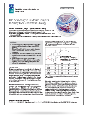
BAs are dichotomized into two principle groups: primary (e.g., cholic acid – CA, chenodeoxycholic acid – CDCA) and secondary (e.g., deoxycholic acid – DCA, lithocholic acid – LCA).1 Primary BAs are synthesized in liver cells (hepatocytes), whereas secondary BAs are produced by an intestinal bacterial-based modification of primary BAs.2 The human liver mainly synthesizes primary bile acids CA and CDCA, while mice mainly synthesize CA and 6-hydroxylated muricholic acids (MCA) from CDCA.3 Bile acids can be further segmented into unconjugated and conjugated (e.g., glycocholic acid – GCA, taurolithocholic acid – TLCA) groups (see Figure 1 for structure summary).
Highlights
• Bile acid composition in ileum and liver was studied using a murine model of microvillus inclusion disease (MVID) in humans.
• CIL’s multiplexed bile acid mixes were used in calibration, quality control standards, and unknown sample preparations.
• Etiology of liver cholestasis caused by the lack of myosin Vb (myoVb) function in human patients with MVID is currently unknown.
• MyoVb knockout in the murine model of MVID demonstrated impact on bile acid levels in ileum and liver suggesting a role for myoVb in regulating hepatic and intestinal bile acid pathways.
Read more by downloading the application note.